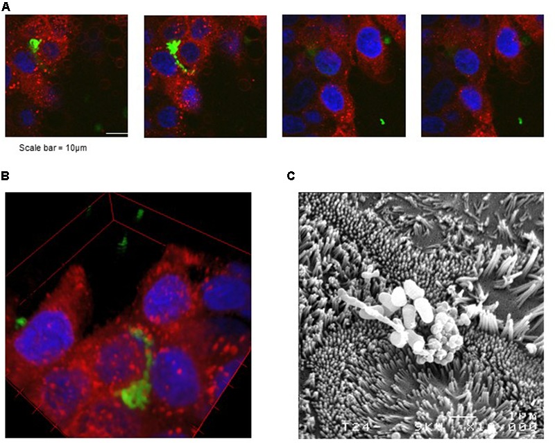
FIGURE 2.

Microscopy imaging of P. freudenreichii Centre International de Ressources Microbiennes–Bactéries d’Intérêt Alimentaire (CIRM-BIA) 129 adhesion to cultured human colon epithelial cells. HT-29 cells were cultured to confluence in DMEM on a slide chamber prior to interaction. P. freudenreichii was cultured in fermented milk ultrafiltrate prior to intracellular labeling of live bacteria using CFSE. Labeled bacteria were then co-incubated with colon cells in a slide chamber prior to washing with PBS and to staining of plasma membrane using Rh-DOPE and mounting in DAPI-containing mounting medium. (A,B) Blue fluorescence evidences colon cells nuclei, red fluorescence their plasma membrane and green fluorescence CFSE-labeled propionibacteria. (A) Z-stack, i.e., confocal images acquired at different “z” altitudes in the labeled preparation. (B) Reconstituted 3-D image showing a cluster of propionibacteria at surface of cells. (C) Scanning electron microscopy observation of propionibacteria adhesion to cultured colon epithelial cells. The same co-incubation was performed in a polycarbonate membrane cell culture insert prior to fixation and scanning electron microscopy observation.
