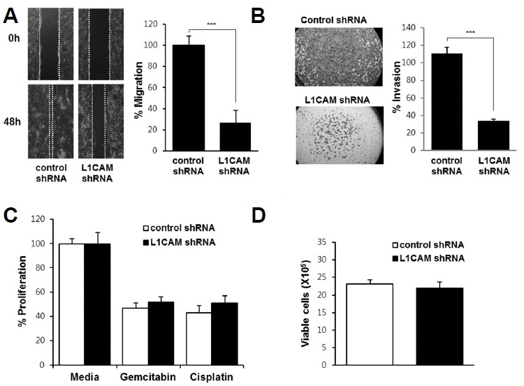
Fig. 3.

Effects of L1CAM knockdown on migration (A), invasion (B), survival (C), and proliferation (D) of EGI-1 cells.
(A) A wound healing assay was performed, with photographs taken immediately after scratching the monolayer (i.e. wound induction) and 48 h later (lower panel). Cell migration was quantified by measuring the distance between the leading edges on either side of the scratch site, plotted as a percentage relative to the zero time point (right panel). (B) A cell invasion assay was performed, with invasive cells quantified by measuring their absorbance at 590 nm. (C) L1CAM-depleted and control cells were treated with 0.5 μg/ml gemcitabin or cisplatin for 72 h and then subjected to the WST-1 cell proliferation assay. (D) L1CAM-depleted and control cells were cultured for 72 h and then cell numbers counted. Three independent experiments were performed in duplicate. Data are expressed as the mean ± SD (***p < 0.001).
