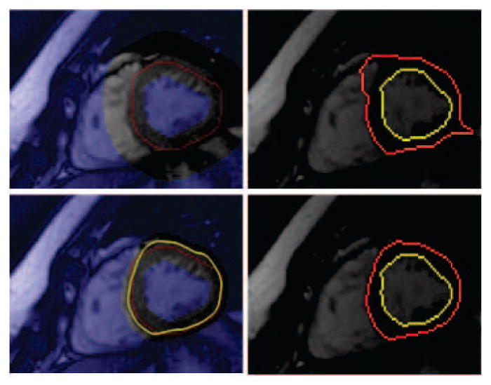
Fig. 3.

Shows the effect of using temporal constraint. In the first row no prior information is used when initializing the and ℬt (left image); whereas on the second row prior epit−1 result (yellow) is used. Images in second column show corresponding segmentation results for the initializations. See text.
