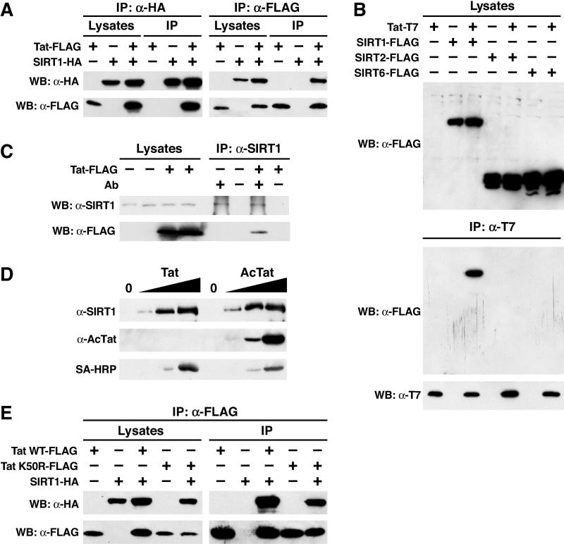
Figure 2. Physical Interaction between Tat and SIRT1.
(A) Immunoprecipitation (IP) and WB of FLAG-tagged Tat (Tat-FLAG) and HA-tagged SIRT1 (SIRT1-HA) after transfection of corresponding expression vectors (+) or empty vector controls (−) into HEK 293 cells.
(B) The same experiments as in (A) performed with T7-tagged Tat and FLAG-tagged SIRT1, SIRT2, and SIRT6.
(C) Coimmunoprecipitation of FLAG-tagged Tat with endogenous SIRT1 in HEK 293 cells transfected with the Tat expression vector or the empty vector control. IPs were performed with or without rabbit α-SIRT1 antibodies.
(D) WB of recombinant SIRT1 protein after pulldown with synthetic biotinylated Tat or AcTat. Tat proteins were detected with antibodies specific for acetylated lysine 50 in the Tat ARM (α-AcTat) or SA-HRP.
(E) Immunoprecipitation/WB of FLAG-tagged Tat or TatK50R and HA-tagged SIRT1. WT, wild type.

