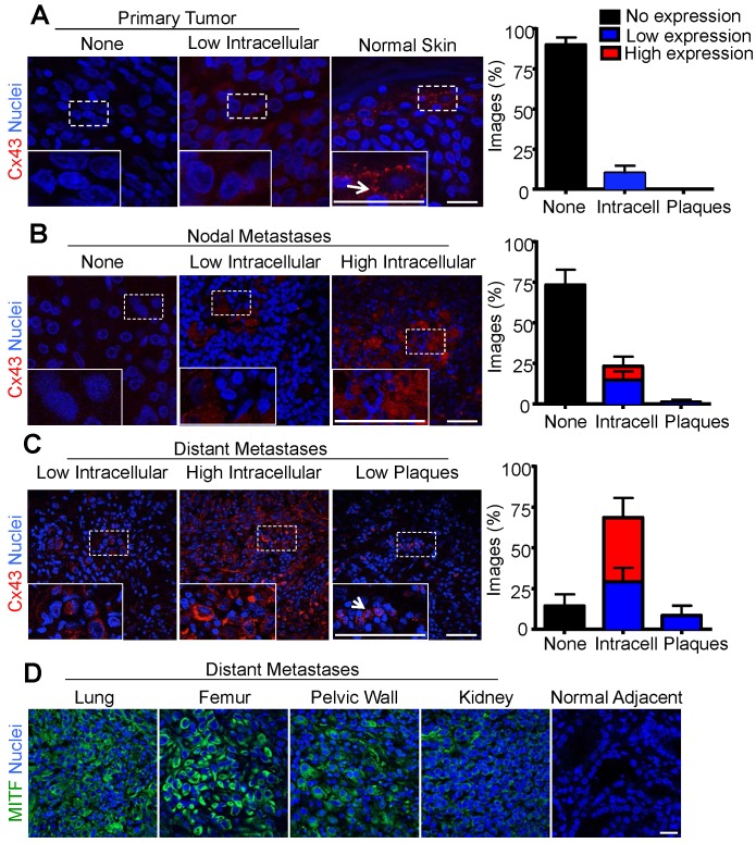
Figure 2.
Increased intracellular Cx43 in distant human melanoma metastases (A) The majority of images analyzed from primary melanoma (N = 14), and (B) nodal metastases (N = 15) did not show evidence of Cx43 expression as opposed to normal skin where Cx43 gap junctions were abundant (A). In contrast, (C) the majority of images analyzed from melanoma metastases to distant organ sites (N = 7) showed evidence of Cx43 expression, however, this expression was predominately intracellular and did not form gap junction plaque-like structures indicative of potentially functional gap junction channels. Arrows indicate punctate Cx43 gap junction structure at the cell-cell interface. (D) Regardless of the distant organ site of melanoma metastases, tumor cores from lung, femur, pelvic wall, and kidney stained positive for MITF, indicating the tumor was from a melanocytic cell lineage. Bars represent mean +/- SEM. Scale bar = 20 μm.

