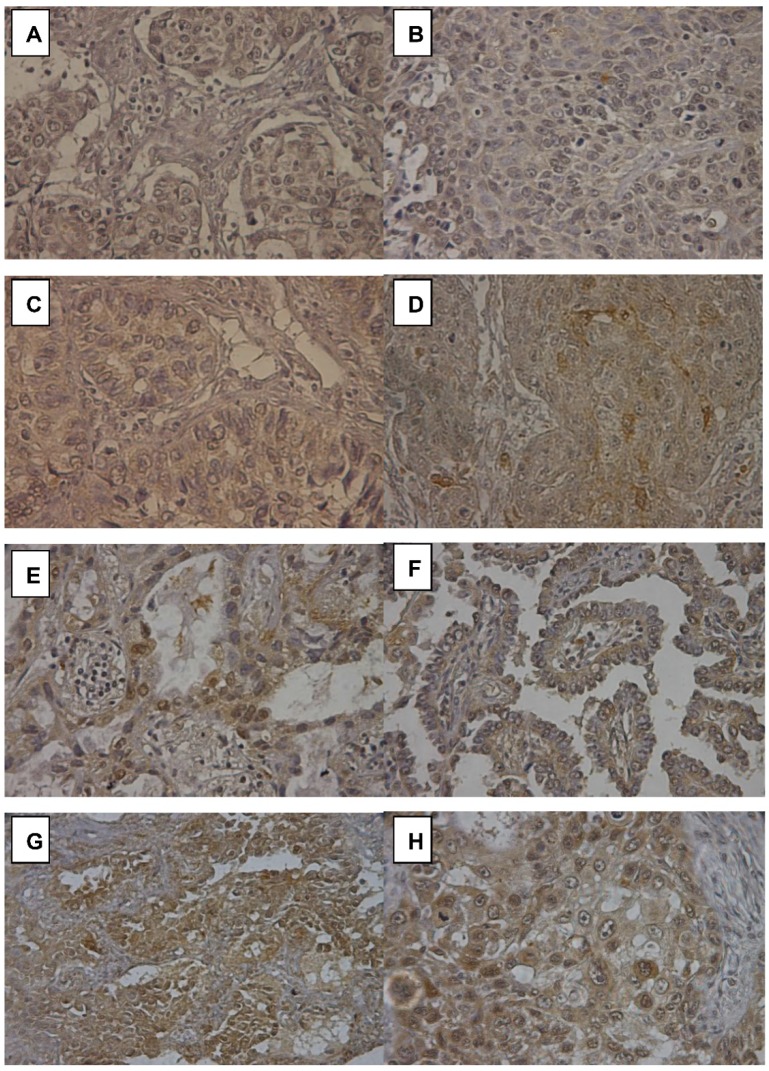
Figure 3.
Immunohistochemistry (IHC) stain of Notch2 in lung adenocarcinoma. (A) and (B) showed negative Notch2 expression. (C) and (D) showed weak Notch2 expression (faint). (E) and (F) showed moderate Notch2 expression. Notch2 was scarcely stained (yellow) in the cytoplasm of tumor cells. (G) and (H) showed high Notch 2 expression (brown) in adenocarcinoma. Notch2 was strongly stained in the cytoplasm of tumor cells. Peripheral normal tissue was a negative control in the same section.

