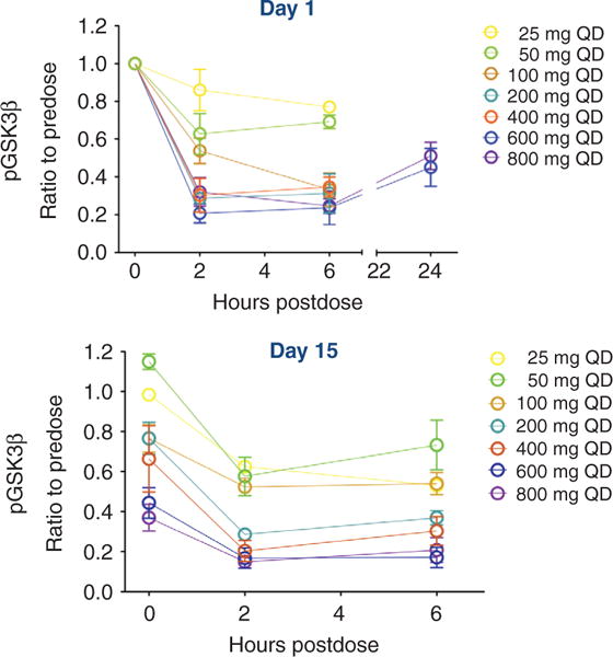
Figure 2.

Ipatasertib shows evidence of PD inhibition of AKT signaling in patients as evaluated by the suppression of pGSK3β in PRP. The ratio of pGSK3β to total GSK3β was evaluated in PRP from patients in all the dosing cohorts (25–800 mg) on day 1 (top) and on day 15 (bottom) of cycle 1 and was graphed as a function of time (in hours) following a single dose of ipatasertib. In all patients, pGSK3β decreased in a time-and dose-dependent manner, reaching a nadir at 2 hours after dose and remaining suppressed at 24 hours after dose. Inhibition of pGSK3β was also sustained at day 15 of dosing (bottom).
