
Fig. 2.
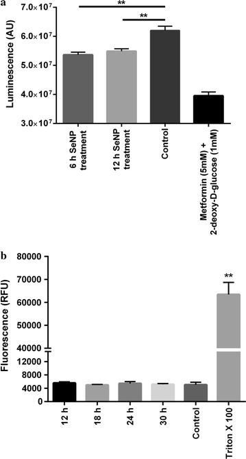
a PC3 cells treated with 2 µg Se/ml SeNPs for 6 or 12 h showed a significant decrease in the levels of cellular ATP compared to the control cells. Metformin and 2-deoxy-d-glucose treated cells were taken as a positive control. ATP was quantified using CellTiter-Glo™ reagent. The intensity of luminescence was proportional to the quantity of ATP present in the sample. The experiment was conducted in triplicate. **p < 0.01 represents a significant difference in the ATP level. b Cells treated with 2 µg Se/ml SeNPs for 6, 12, 18, 24, or 30 h showed no LDH release in the culture medium compared to the negative control (PBS treated cells). Triton-X 100 treated cells were taken as a positive control. The intensity of fluorescence was directly proportional to the quantity of LDH present in the sample. The experiment was conducted in triplicate. **p < 0.01 represents a significant difference in the LDH levels of positive control and SeNP treated cells