
Figure 2.
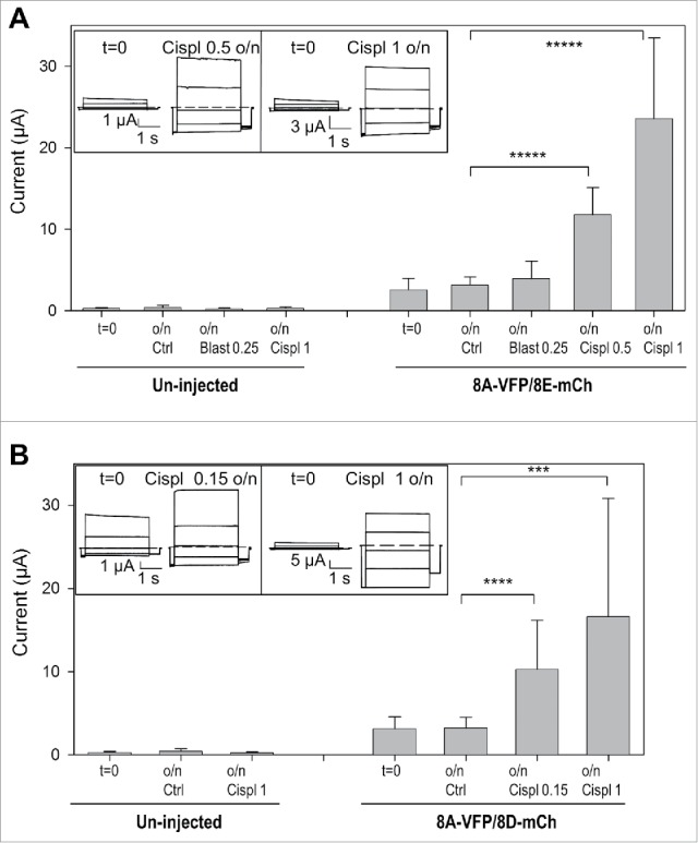
Cisplatin incubation strongly increases currents of oocytes expressing 8A-VFP/8E-mCh or 8A-VFP/8D-mCh. (A) Mean currents from un-injected or from 8A-VFP/8E-mCh injected oocytes measured before (t = 0; un-injected n = 29; 8A-VFP/8E-mCh n = 60) and after overnight incubation in “Maintaining” solution (Ctrl; un-injected n = 11; 8A-VFP/8E-mCh n = 29), 0.25 mM blasticidin (Blast 0.25; un-injected n = 4; 8A-VFP/8E-mCh n = 9), 0.5 mM cisplatin (Cispl 0.5; 8A-VFP/8E-mCh n = 7) or 1 mM cisplatin (Cispl 1; un-injected n = 14; 8A-VFP/8E-mCh n = 15; *****P < 10−9). The insets show 8A-VFP/8E-mCh traces from one oocyte before and after overnight incubation in 0.5 mM (left) or 1 mM cisplatin (right). (B) Bars represent currents of un-injected or 8A-VFP/8D-mCh injected oocytes recorded before (t = 0; un-injected n = 19; 8A-VFP/8D-mCh n = 39) and after overnight incubation in “Maintaining” solution (Ctrl; un-injected n = 8; 8A-VFP/8D-mCh n = 21), 0.15 mM cisplatin (Cispl 0.15; 8A-VFP/8D-mCh n = 6) or 1 mM cisplatin (Cispl 1; un-injected n = 11; 8A-VFP/8D-mCh n = 12; ***P < 0.001; ****P < 0.0001). The insets show typical 8A-VFP/8D-mCh traces from one oocyte before and after overnight incubation in 0.15 mM (left) or 1 mM cisplatin (right). Error bars indicate SD.