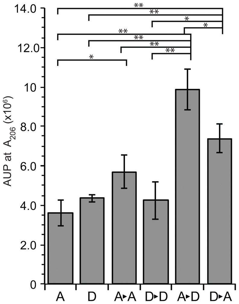
Figure 2.

Liberated PG monomers from sequential digestion of sacculi with LtgA and LtgD. Soluble PG fragments from whole sacculi digests with LtgA or LtgD followed by boiling and a subsequent digest with LtgA or LtgD were separated by HPLC. The abundance of PG monomers (tripeptide (GaM-3) and tetrapeptide (GaM-4)) was determined by calculating the peak area (AUP) from three independent experiments. Horizontal bars indicate significance of P < 0.05, and (*) indicates P < 0.01 by Student’s t-test. Error bars represent standard deviation.
