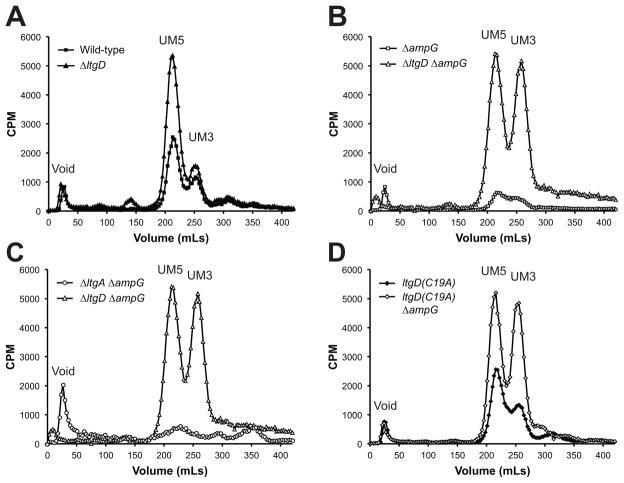
Figure 6.
Cellular PG fragments extracted from LT and ampG mutants. Hot water extracts from cultures labeled with [3H]-glucosamine were separated by size-exclusion chromatography to determine the abundance of PG precursors and fragments in the cell. (A) Mutation of ltgD resulted in increased cellular UDP-MurNAc-pentapeptide compared to wild-type. (B) Mutation of ampG decreased the amount of detectable PG precursors, but an ltgD ampG mutant increased UDP-MurNAc-pentapeptide and UDP-MurNAc-tripeptide compared to wild-type. (C) A double ltgA ampG mutant resulted in a decrease in the amount of radiolabeled PG fragments in the cell. (D) The ltgD(C19A) lipidation mutant contained wild-type levels of PG precursors, but the ltgD(C19A) ampG mutant contained high levels of UDP-MurNAc-tripeptide and -pentapeptide like the ltgD ampG mutant. UM5, UDP-MurNAc-pentapeptide; UM3, UDP-MurNAc-tripeptide.

