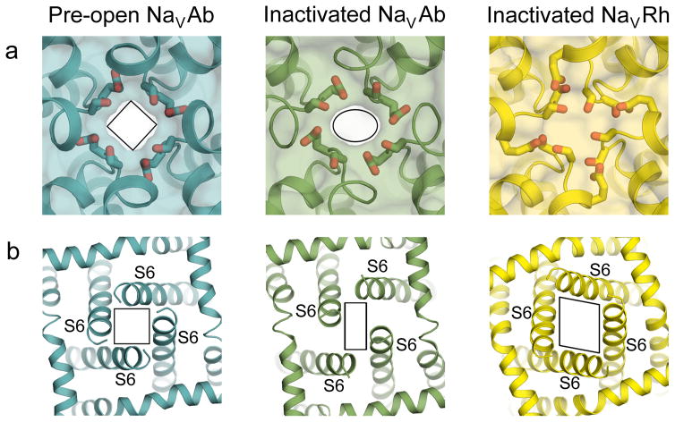
Figure 6. Conformational changes in the pore associated with slow inactivation.
a, An extracellular view of the pore through the selectivity filter. The selectivity filter collapses from a four-fold symmetric shape in pre-open NaVAb (left) to an oval shape in inactivated NaVAb (middle) to a completely closed pore in inactivated NaVRh (right). b, An intracellular view of the pore at the C-termini of the S6 segments. The pore distorts from a square shape in the pre-open NaVAb structure to a parallelogram shape in the inactivated NaVAb and NaVRh structures.

