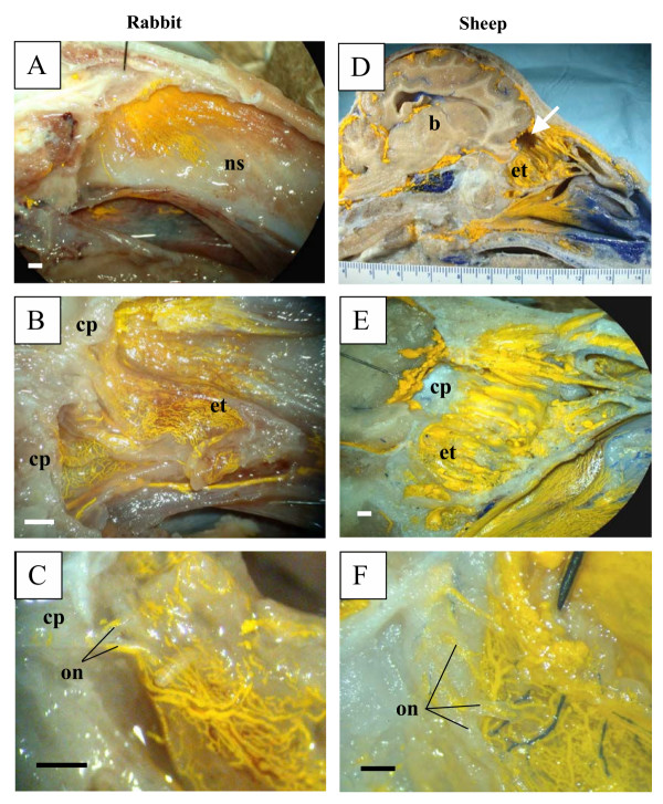
Figure 2.
Microfil distribution patterns in the head of rabbits and sheep. All images are presented in sagittal plane with gradual magnification of the olfactory area adjacent to the cribriform plate. Reference scales are provided either as a ruler in the image (mm) or as a longitudinal bar (1 mm). A-C illustrates images of the rabbit and D-F images of sheep. In both species, the Microfil can be viewed in the perineurial spaces of the olfactory nerves external to the cranium, which merge into network of lymphatic vessels in the ethmoid turbinal systems. b – brain; cp – cribriform plate; et – ethmoid turbinates; on – olfactory nerves; ns – nasal septum; arrow in D – portion of cribriform plate removed for histology.

