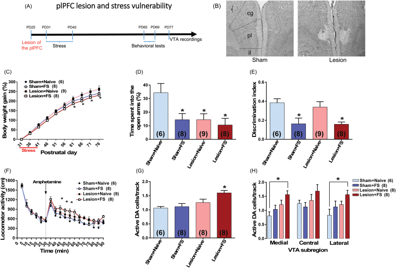
Fig. 2.
plPFC disruption increases vulnerability to adolescent stress in adult rats. (A) plPFC lesion was induced in rats at PD25. Six days after surgery, adolescent rats (n = 6–9/group) were submitted to footshock (FS). At adulthood, they were tested in the elevated plus-maze (EPM) (PD65), novel-object recognition (NOR) test (PD66–67), locomotor response to amphetamine (PD68) and for ventral tegmental area (VTA) dopamine (DA) cells activity (PD77–94). (B) Photomicrographs show the adolescent plPFC lesion in an adult animal. The same region is also shown in sham animals; cg, cingulate PFC; pl, plPFC; il, infralimbic PFC. (C) FS exposure induced impairment in body weight gain. (D) FS by itself also induced anxiety-like responses in the EPM and (E) a cognitive impairment in the NOR test. Similar changes were observed in animals with plPFC lesion and exposed to FS. The lesion by itself induced an anxiety-like response but did not induce any change in the NOR test. (F) plPFC lesioned animals exposed to FS also showed an enhanced locomotor response to amphetamine (0.5 mg/kg; injection is indicated by the dashed line) and (G) an increased VTA DA population activity. (H) This increased DA population activity was observed throughout the VTA with significant differences in both medial and lateral VTA. In the VTA recordings, data from 1 plPFC lesion + naïve animal were excluded due to electrode misplacement. Data are presented as mean ± SEM. *P < .05 vs naive rats.

