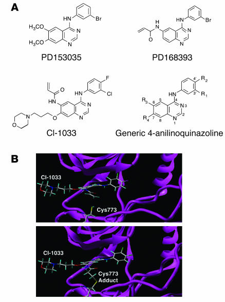
Figure 1.
Structure of ErbB tyrosine kinase inhibitors. (A) Chemical structures of ErbB tyrosine kinase inhibitors. (B) Molecular model of CI-1033 interaction with the ErbB-1 kinase domain. The upper panel shows the model of the noncovalently bound CI-1033 in the ATP-binding site of the tyrosine kinase ErbB-1 (protein data bank: 1M17) crystal structure. The Tarceva compound was removed, and CI-1033 was manually modeled into the site. The backbone of the kinase is shown as a ribbon diagram along with the atoms of Cys773. The lower panel shows the model of the covalently bound CI-1033 in the ATP-binding site. The atoms are shown as stick models with carbon atoms colored white, oxygen atoms red, nitrogen atoms dark blue, sulfur atoms yellow, and hydrogen atoms light blue.

