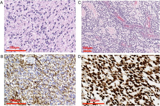
Fig. 2.
Canine glioma histology. (A) Anaplastic astrocytoma with pleomorphism, anisokaryosis, nuclear atypia, and mitotic activity. (B) GFAP staining of anaplastic astrocytoma. (C) High-grade oligodendroglioma composed of ovoid to fusiform cells and endothelial vascular proliferation. (D) Olig2 immunohistochemistry demonstrates strong nuclear positivity of more than 90% of neoplastic cells. Images courtesy of Dr Miller, Purdue Veterinary Medicine.

