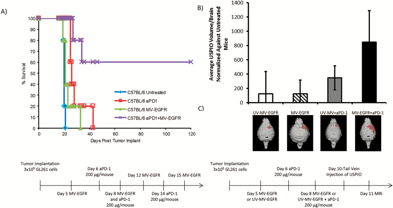
Fig. 3.
MV-EGFR+aPD-1 therapy significantly enhanced survival of mice bearing orthotopic GL261 gliomas and increased T cell influx into their brains as assessed by T2* MRI. (A) Kaplan‒Meier survival curves of mice bearing orthotopic GL261 gliomas treated as indicated. MV-EGFR+aPD-1 treatment resulted in significant enhancement of survival compared with all other groups. aPD-1 therapy resulted in significant enhancement of survival compared with untreated mice but not compared with MV-EGFR treated mice. (B) T2* MRI analysis of T-cell influx into mouse brains on day 11 post tumor implantation. MV-EGFR+aPD-1 therapy group had increased T-cell influx compared with other groups. (C) Representative T2* MRI reconstructions (red shading = USPIO positive).

