Fig. 4.
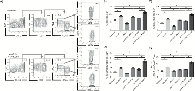
MV-EGFR+aPD-1 treatment enhances CD8 T-cell influx into the brain of GL261-bearing animals. Brains isolated from treated GL261-bearing animals were assessed for immune cell infiltration (N=9 per group) (A) Representative flow cytometry histograms depicting gating scheme. (B) An increased proportion of T cells (CD3+CD45.2hi) were isolated from animals treated with combination therapy compared with monotherapy. (C) A higher proportion of CD8 T cells (CD8α+CD3+CD45.2hi) were isolated from the brains of dually treated animals, and (D) more of those cells were positive for granzyme B (GzmB+). (E) An increase in the CD8 T cell:Foxp3+ Treg ratio in the brain was also observed in MV-EGFR+aPD-1 treated animals. Error bars represent mean±SEM. * denotes P<.05.

