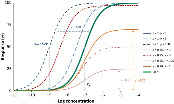
Figure 4.

Response curves with the present model (eq. 31) for a ligand of 100 nmol/L affinity (K d = 10–7 mol/L) for a full agonist (ε = 1, blue lines) at different amplifications (γ = 1, 3, and 100) and a weak partial agonist (ε = 0.25, red lines) at the same amplifications (γ = 1, 3, and 100). Another partial agonist without amplification is also included (ε = 0.70, γ = 1 orange line) for comparison. Note that with the present model, the basic parametrization (ε = 1, γ = 1) fully reproduces the Clark model (blue and double green lines completely overlap), which could not be done with the previous models (Figure 2). The effect of different post‐activation amplifications on the observed response of a full and a partial agonist is also illustrated in more detail in Figure 6.
