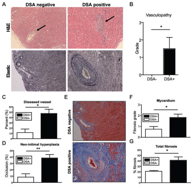
Figure 3. Histological analysis of representative graft tissue of DSA+ and DSA– recipients.
(A) Representative sections of explanted graft at day 100 from DSA+ and DSA– recipients were stained with H&E and elastic staining for evaluation of CAV. Note the significant vasculopathy (arrow) in the DSA+ recipients and, in contrast, the relatively normal vascular integrity in the DSA– recipients at 100 days after transplantation. Original magnification ×200. (B) Vasculopathy was scored on elastic stains as 0, none; 1, mild, 0–10% of the luminal area compromised; 2, moderate, 10–50% of the luminal area compromised and 3, severe, greater than 50% of the luminal area compromised. Higher degrees of vasculopathy were identified in the graft from DSA+ recipients compared to DSA– recipients. (C) Morphometric analysis demonstrated more narrowing of vascular lumina in DSA+ recipients 100 days after transplantation than DSA–recipients. (D) Vessels showing more than 20% of occlusion were counted as diseased vessels. DSA+ recipients showed increased number of diseased vessels at day 100. (E) Representative of trichrome stained grafts showed the diffuse fibrosis and thickened vascular wall (stained blue), consistent with CAV. (F) Fibrosis was semi-quantitatively scored in the myocardium as 0, none; 1, 0–10%; 2, 10–50% and 3, >50% of the myocardial area. Increased level of fibrosis in myocardial area of DSA+ recipients was identified compared to DSA–recipients. Original magnification ×200. (G) Total fibrosis measured from entire explanted graft showed significantly increased in DSA+ recipients compared to DSA– recipients. The percentage of total fibrosis was quantified and expressed as% positive trichrome pixels. Data here are expressed as mean ± SEM of six grafts in each group. *p < 0.05, **p < 0.01.

