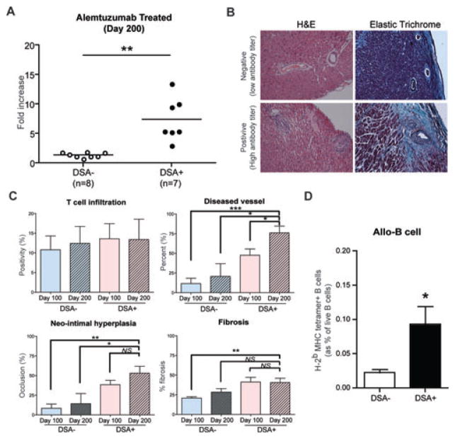
Figure 7. Alloantibody production and chronic rejection development in alemtuzumab treated CD52Tg recipients at day 200.
(A) Serum alloantibody detection with flow crossmatch showed two distinct phenotypes either with low alloantibody titer (negative) or with high alloantibody titer (positive). (B) Representative sections of explanted graft at day 200 from DSA+ and DSA– recipients were stained with H&E and elastic trichrome staining for evaluation of CAV. Original magnification ×200. (C) The quantification of T cell infiltration showed similar level of intragraft T cell infiltration in all groups. DSA+ recipients showed increased number of diseased vessel at day 200. Mophometric analysis demonstrated only a trend of increased neo-intimal hyperplasia in DSA+ recipients at 200 days after transplantation than DSA– recipients. Total fibrosis measured from entire explanted graft showed no difference in DSA+ recipients compared to DSA– recipients. Data are expressed as mean ± SEM of six grafts in each group. (D) The DSA+ recipients (n = 7) showed significantly increased levels of alloreactive (anti-Kb/Db) B cells in the spleen compared to DSA– recipients (n = 8). NS > 0.05, *p < 0.05, **p < 0.01, ***p < 0.001.

