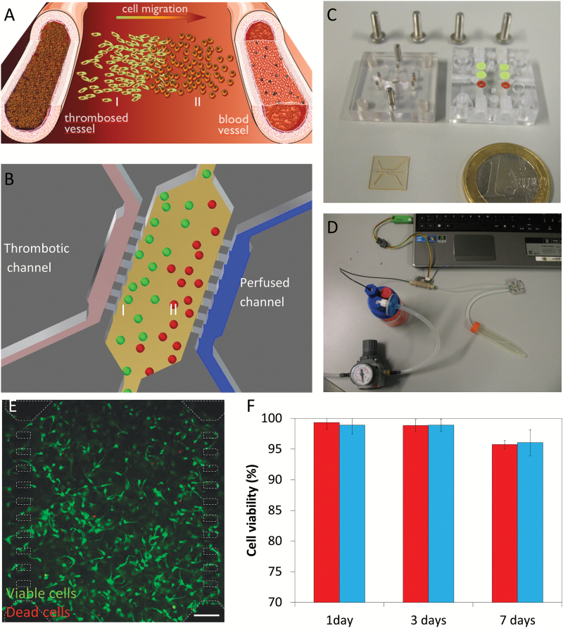
Fig. 1.
Experimental setup. (A) Scheme of pseudopalisade formation. Under obstructed conditions, nutrient scarcity triggers a migratory response in those cells located in the obstructed blood vessel vicinity (I) towards enriched regions (II). (B) Experimental scheme within the microdevice mimicking the obstructed conditions and the starved (I) and enriched (II) regions. (C) Fabricated microdevice and packaging tool. (D) Microfluidic system. (E) U-251 cell viability within the microdevice after 9 days, live cells (labeled with calcein 1 µg/ml) are shown in green, whereas dead cells are shown in red (labeled with propidium iodide 4 µg/ml). Microdevice posts (50x100 µm) are delimited in white dashed line. Cells were cultured at 4 million cells/ml within a 1.5 mg/ml collagen hydrogel. Viable cells are shown in green, whereas dead cells are shown in red. (F) Comparison of cell viability between hydrogels on Petri dishes (red) and within the microdevice (blue). Cell viability is expressed as the percentage of live cells. Scale bar is 200 µm.

