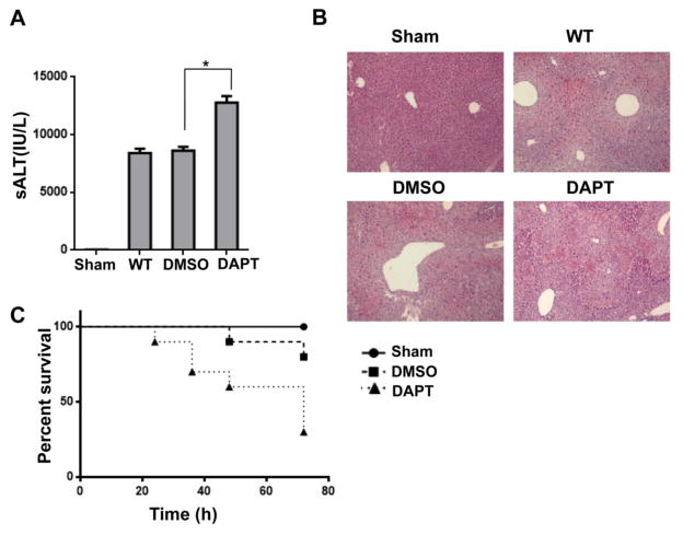
Figure 1. Blockade of Notch signaling pathway aggravates APAP-induced liver damage.
Mice were subjected to injection of γ-secretase inhibitor DAPT (10 mg/kg, Sigma-Aldrich, MO) or DMSO vehicle via tail vein at 30 min prior to APAP (400 mg/kg) challenge. (A) Hepatocellular function, assessed by serum ALT levels (IU/L). Results expressed as mean±SD (n=4–6 samples/group), *p<0.01. (B) Representative histological staining (H&E) of APAP-conditioning liver tissue (n=4–6/group). Original magnification x100. (C) Animal survival curves after a single dose of APAP treatment over 72 hours (p< 0.05).

