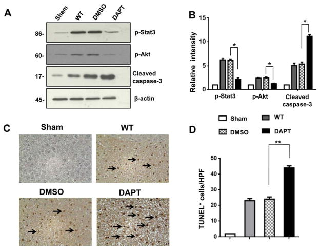
Figure 4. Blockade of Notch signaling pathway promotes APAP-induced hepatocellular apoptosis.
(A) Western blot analysis of phos-Stat3, phos-Akt, and cleaved caspase-3 in APAP-challenged livers after DAPT or DMSO treatment. β-actin served as an internal control. Data representative of three experiments. (B) Density ratios of Hes1, HMGB1, TLR4, and NLRP3. *p<0.05. (C) TUNEL-assisted detection of apoptosis in APAP-challenged liver tissue. Representative of 4–6 mice/group. (D) TUNEL+ cells were scored semi-quantitatively by averaging the number of apoptotic cells (mean±SD) per field at 200× magnification. A minimum of ten fields was evaluated per sample. *p<0.01. Representative of 4–6 mice/group.

