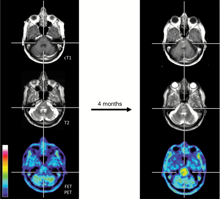
Fig. 3.
Seventy-three-year-old patient (patient ID #24) with a rapid progressive tumor of the brainstem (histology of this brainstem lesion could not be obtained due to the patient’s refusal). In correspondence to the clinical deterioration (ie, dysphagia, dysarthria) within 4 months, a contrast-enhancing lesion, a progression of the T2 signal, and an increase of the metabolic activity (TBRmax baseline, 1.6; TBRmax follow-up, 3.2) compared with baseline imaging (left column) are illustrated.

