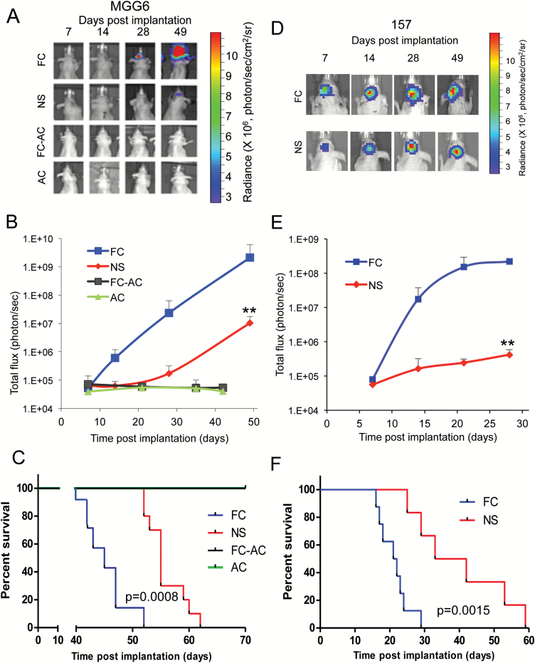
Fig. 3.
Floating cells form aggressive tumors in vivo. (A–C) Athymic mice were implanted with ~50000 of either FC, NS, FC-AC, or AC from MGG6 expressing Fluc into the left forebrain (n = 7–10). Representative pseudo-color images of Fluc bioluminescence at different time point post-implantation (A). Quantification of Fluc radiance intensity presented as photons/sec/cm2/surface radiance; data presented as mean±SD, n = 10; **P < .01 FC vs NS by ANOVA and Tukey’s post-hoc test (B). Kaplan–Meier survival curves; n = 7–10; ** P < .01 FC vs NS by Mantel–Cox (log-rank) test (C). (D–F) Athymic mice were implanted with ~50000 NS or FC cells from GSCs 157 and results were analyzed similar to A–C; n = 8; ** P < .01 FC vs NS.

