Fig. 4.
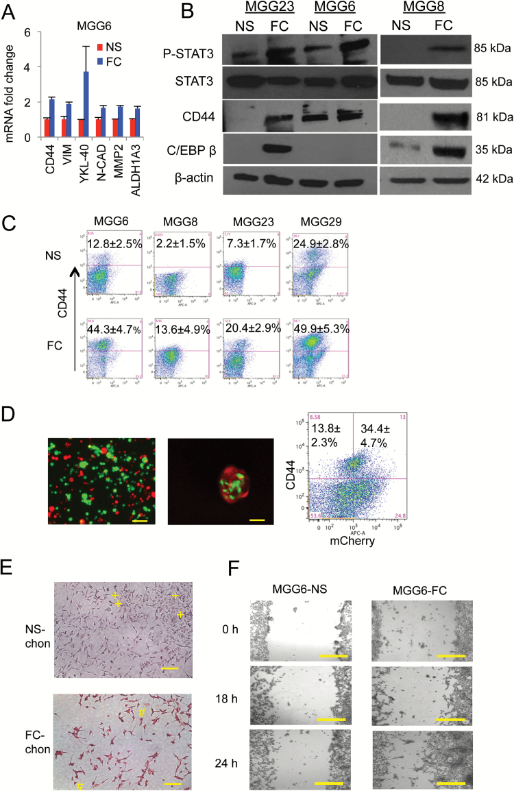
Floating cells display enhanced mesenchymal properties. (A) MGG6 NS and FC mRNA were analyzed by qRT-PCR for different mesenchymal markers including CD44, vimentin, YKL-40, N-cad, and MMP2. (B) Cell lysates from different cultures were analyzed by western blotting for pSTAT3, STAT3, CD44, C/EBPβ, and β-actin. (C) FACS analysis showing proportion of CD44 positive cells in FC and their corresponding NS from different GSC cultures. (D) MGG29 NS were fractionated into CD44high and CD44low subpopulation by FACS and predominant cells were established and labeled with GFP and mCherry, respectively. Left: fluorescence analysis of a mixture of these 2 subpopulations cultured in serum; middle: FC collected and analyzed for both GFP and mCherry; right: flow cytometry of CD44high cells in both GFP and mCherry populations. (E) NS and FC were differentiated using StemPro Chondrogenesis Differentiation Kit and stained with fast green and safranin O (positive, #; negative, +). (F) NS and FC were cultured as a monolayer and their ability to invade/migrate was evaluated using scratch healing assay. Bright field micrographs are showing for different groups. Scale bar, 50 μm.

