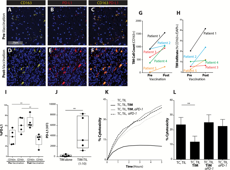
Fig. 1.
GBM TIMs expand to inhibit vaccine-induced T-cell mediated tumor cytolysis via PD-1/PD-L1 regulatory pathway. (A, D) CD163, DAPI, (B, E) PD-L1, DAPI, and (C, F) CD163, PD-L1, and DAPI co-staining are shown across pre- and post-DC vaccination samples from a GBM patient at 40× magnification (scale bar represents 75µm). (G) CD163+ cell count across pre– and post–DC vaccine treatment patient samples was quantified (n = 5). (H) Percent of CD163+ cells of total number of cells (DAPI+) was quantified (n = 5). (I) The percent of CD163+ cells dually expressing PD-L1 was quantified before and after vaccination (n = 5) (**P < .01). (J) PD-L1 expression on CD11b+ TIMs in the absence or presence of CD3+ TILs from freshly resected GBM shown (**P < .01) (n = 5). (K) Representative plot demonstrating TIL cytolysis of tumor cells (TC) over time in the absence or presence of TIMs or PD-1 mAb shown for freshly resected GBM. (L) GBM tumor cell cytolysis at selected time point of 4 hours in the absence or presence of TIMs or PD-1 mAb shown (n = 11/group) (**P < .01).

