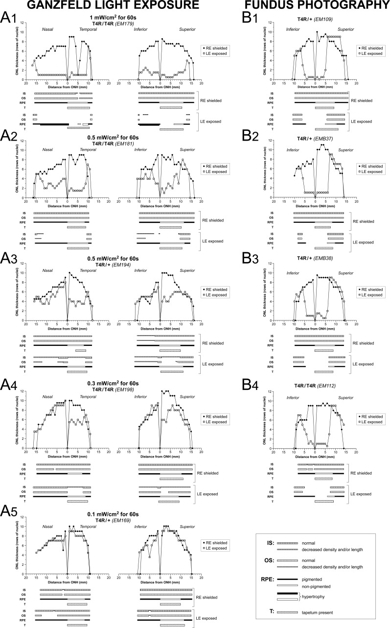
Figure 4.
Morphometric analysis of retinal changes 2 weeks after light exposure. Spider graphs of ONL thickness and schematic representation of IS, OS, and RPE structure 2 weeks post light exposure of the LE of RHO T4R mutant dogs. The RE was shielded. Data derived from retinal histologic sections extending from the edge of the optic nerve head to the superior, inferior, nasal, and temporal ora serrata. T, tapetum.

