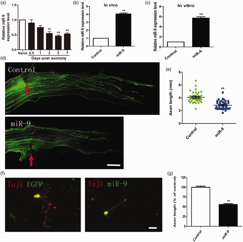
Figure 1.
MiR-9 overexpression inhibited axon regeneration in adult sensory neurons in vitro and in vivo. (a) qRT-PCR data indicating miR-9 levels in adult dorsal root ganglions after sciatic nerve injury. Note that miR-9 expression was significantly down-regulated from three to seven days after sciatic nerve axotomy (n = 3 for each condition). Error bars represent SEM. **P < 0.01. (b) and (c) Quantification of miR-9 mRNA level at three days after electroporation of miR-9 mimics in vivo and in vitro (n = 3 for each condition). Error bars represent SEM. **P < 0.01. (d) Representative images of EGFP-labeled regenerating axons in the whole-mount sciatic nerves. Red arrowheads mark the crush sites. Bar = 500 µm. (e) Scatter plot of average lengths of regenerating sciatic nerve axons (n = 6 mice for each condition). Error bars represent SEM. **P < 0.01. (f) Representative images of cultured adult mouse sensory neurons expressing EGFP (green), EGFP + miR-9 mimics (miR-9). All neurons were stained with Tuj1 (red). Scale bar = 100 µm. (g) Quantification of the average length of the longest axons (normalized to the average length of the control axons, n = 3). Error bars represent SEM. **P < 0.01.

