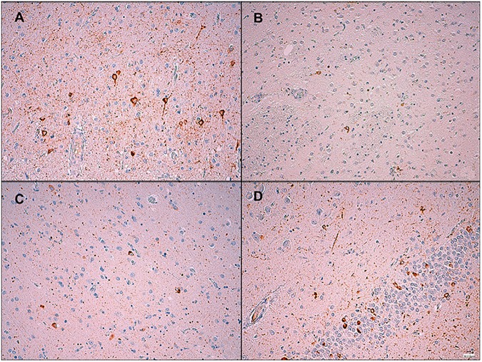
Fig 3. Tau immunohistochemistry from a GRN+/A152T+ patient diagnosed with unclassifiable tauopathy.
(A) Strong diffuse neuronal cytoplasm immunoreactivity and threads in the cerebral cortex, x200. (B) Mild immunoreactivity in the neuronal cytoplasm and processes in the basal ganglia, x200. (C) Moderate diffuse neuronal cytoplasm immunoreactivity, some neurofibrillary tangles and grains in the neuronal processes in the amygdala, x200. (D) Moderate diffuse neuronal cytoplasm immunoreactivity in the neuronal cytoplasm of the dentate gyrus and threads in the hippocampus, x200.

