Figure 5.
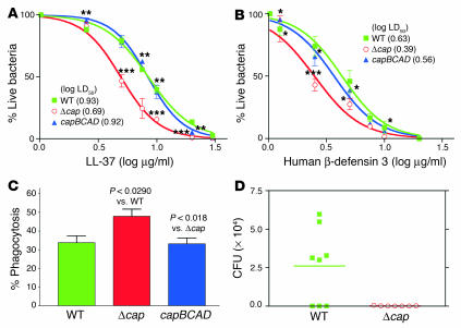
Role of PGA in immune evasion and virulence of S. epidermidis. (A and B) Resistance to cationic antimicrobial peptides. Washed S. epidermidis cells (approximately 105) were incubated with LL-37 (A) or human β-defensin 3 (B) in various concentrations for 2 hours at 37°C. Thereafter, S. epidermidis survivor cells were counted by plating. Results are shown as dose-response curves. The log LD50 values for all strain/peptide combinations are given in the key. Statistical analyses are for each peptide concentration. Values of significance were calculated against the wild-type (for Δcap) and Δcap (for capBCAD) strains. (C) Resistance to neutrophil phagocytosis. Phagocytosis by human neutrophils was determined after 30 minutes of incubation with S. epidermidis at a ratio of 20 bacteria per PMN. (D) Mouse model of subcutaneous device-related infection. Catheter pieces with equal amounts of adhered S. epidermidis cells (2 × 10⁵) were placed under the dorsum of the animals. CFU on implanted devices 1 week after infection were counted. The horizontal bar shows the group mean. *P < 0.05; ***P < 0.01; ***P < 0.001.

