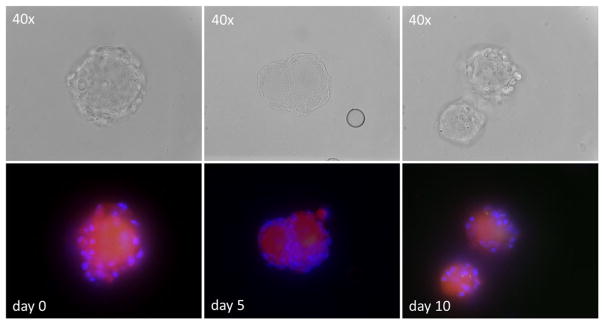
Fig. 1.
Viability and morphology of dissociated human trigeminal ganglia (TG) in culture. Mechanically dissociated human TG were cultured in six-well glass plates for the indicated days. Viable neurons incorporate the red-fluorescent vital dye with accompanying non-neuronal, probably satellite cell nuclei identified by DAPI (blue). Top row shows the same viable neurons seen in phase-contrast microscopy

