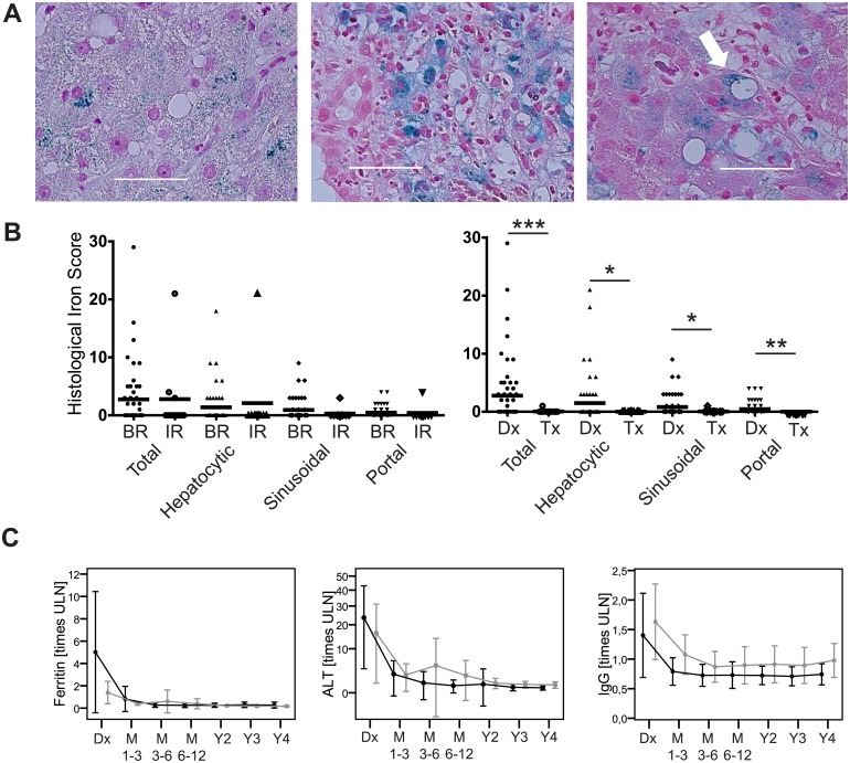
Fig 2. Reversible hyperferritinemia and mild iron deposition in untreated AIH-1.
(A) Intrahepatic iron deposition (blue granula) in untreated AIH-1 in (left) hepatocytes, (middle) portal fields and (right) the sinusoidal compartment (white arrow). (B) The semi-quantitative histopathological iron deposition score (left) in untreated AIH-1 with subsequent biochemical remission (BR: N = 47) or incomplete response (IR: N = 10) and (right) under therapy (Tx; N = 24) compared to baseline at diagnosis (Dx; N = 61). (C) Longitudinal course (mean and standard deviation) under therapy (M = month; Y = year) in BR (black) and IR (grey). (* p<0.05; ** p<0.01; *** p<0.001; ULN = upper limit of normal).

