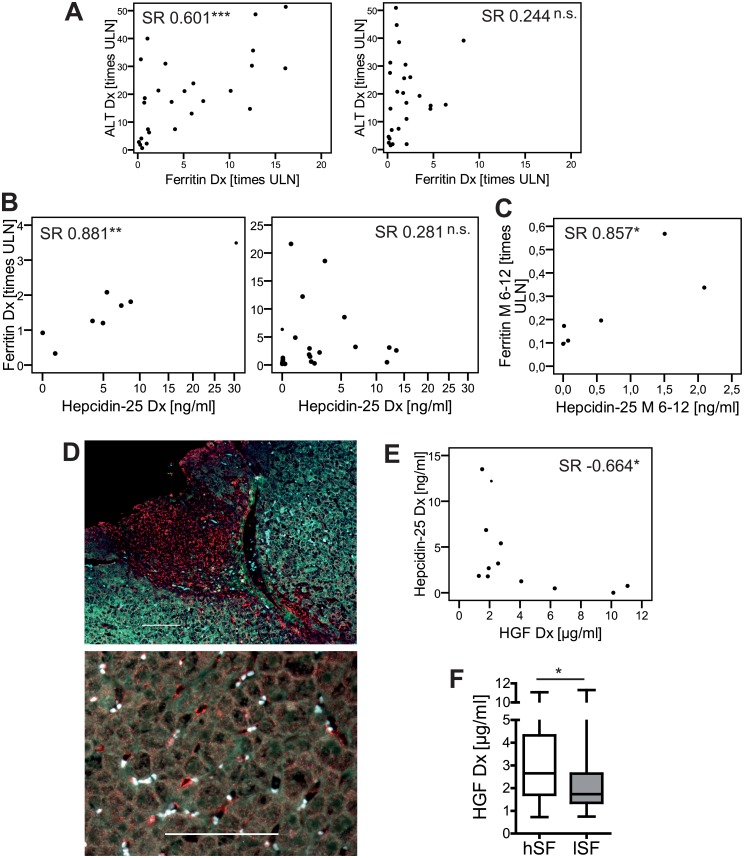
Fig 3. Iron homeostasis is potentially deregulated by HGF driven suppression of hepcidin-25 in untreated AIH-1.
(A) Spearman rank correlation (SR) analysis of serum ferritin in AIH-1 with alanine aminotransferase (ALT) at diagnosis (Dx) in patients with subsequent biochemical remission (BR, left panel, N = 24) and incomplete biochemical response (IR, right panel, N = 24) matched for ALT, gender and age as far as possible. (B) SR analysis of serum ferritin and hepcidin-25 in patients with IR (N = 8; left panel) and with BR (N = 21; right panel) upon standard therapy and (C) in patients with achieved BR after 6–12 months of therapy (M 6–12; right; N = 7). (D) The hepatocyte growth factor (HGF, red; autofluorescence in green and blue) is expressed in (top) the portal tracts, endothelium and (bottom) liver sinusoids in a representative liver biopsy of untreated AIH-1. White bars represent 100 μm. (E) SR analysis of HGF and hepcidin-25 in untreated AIH-1 (N = 12) with subsequent BR and high ferritin (>2,09x ULN). (F) HGF in patients with high (hSF, N = 30) and low serum ferritin (lSF, N = 37). (* p<0.05; ** p<0.01; *** p<0.001; not significant, p≥0.05; ULN = upper limit of normal)

