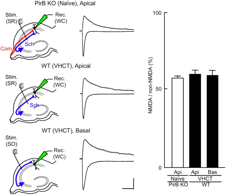
Fig 5. Effect of PirB deficiency on the amplitude of NMDA EPSCs evoked in CA1 pyramidal neuron synapses.
Schematic diagrams showing the arrangement of electrodes for recording. A stimulating electrode was placed in the stratum radiatum [Stim. (SR)] or the stratum oriens [Stim. (SO)] of the CA1 to activate apical or basal synapses, respectively. Whole-cell patch recordings [Rec. (WC)] were made from CA1 pyramidal neurons. Sample superimposed traces of representative EPSCs recorded in hippocampal slices prepared from PirB KO and WT mice. The top traces show NMDA EPSCs at +30 mV in the presence of DNQX and bicuculline. The bottom traces show non-NMDA EPSCs at −90 mV in the presence of bicuculline. Each trace was averaged from five consecutive recordings. Scale bars: 50 pA (vertical) and 100 ms (horizontal). Relative amplitudes of NMDA EPSCs are expressed as percentages of non-NMDA EPSCs. Columns and error bars represent means and SEM, respectively (n = 7 each; P > 0.05, t-test). Api, apical synapses; Bas, basal synapses.

