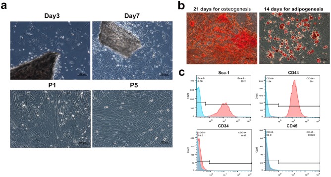
Fig 1. Characteristics and identification of compact bone-derived mouse mesenchymal stem cells.
(a) Spindle cells migrated out from bone chips after a 3-d culture and reached 80–90% confluence after additional 4 days. A monolayer of homogeneous vortex-shaped cells were observed on passage 5. (b) Mineralized nodule were assayed by alizarin red staining and adipogenesis of MSC was stained with Oil red O. (c) Flow cytometric analysis showed that the cells were positive for Sca-1 and CD44 and negative for CD34 and CD45.

