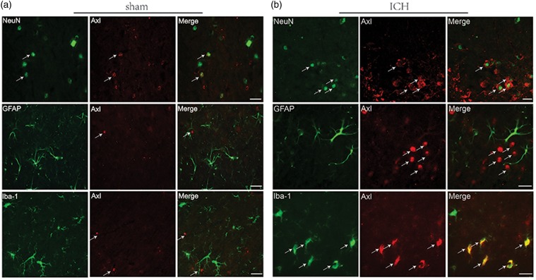
Figure 2.
Endogenous Axl preferentially expressed on cellular membrane of neuron and microglia. Representative images of immunofluorescent staining to show the expression profile both in (a) sham and (b) ICH mice brain of Axl (red), respectively, with NeuN (green) marked neurons, GFAP (green) marked astrocytes and Iba-1 (green) marked microglia. Samples were obtained from peri-hematoma area 24 h following autologous blood-injection-induced ICH. Bar=20 µm.

