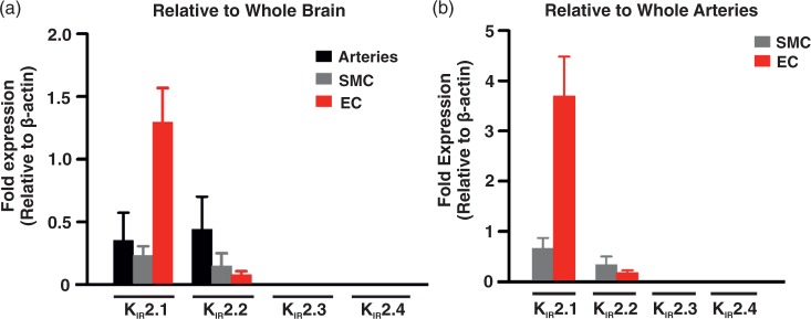
Figure 5.
Q-PCR analysis of KIR 2.x subtypes. mRNA expression (a) of KIR 2.1–2.4 in whole middle cerebral arteries (n = 4), isolated smooth muscle (n = 4), and endothelial (n = 4) cells relative to whole brain mRNA expression. mRNA expression (b) of KIR 2.1–2.4 in isolated SMCs (n = 4) and ECs (n = 4) cells relative to whole middle cerebral arteries mRNA expression. Data (means ± SE) are expressed relative to β-actin expression. Note that KIR 2.3 and 2.4 mRNA was not detectable in selected tissues.

