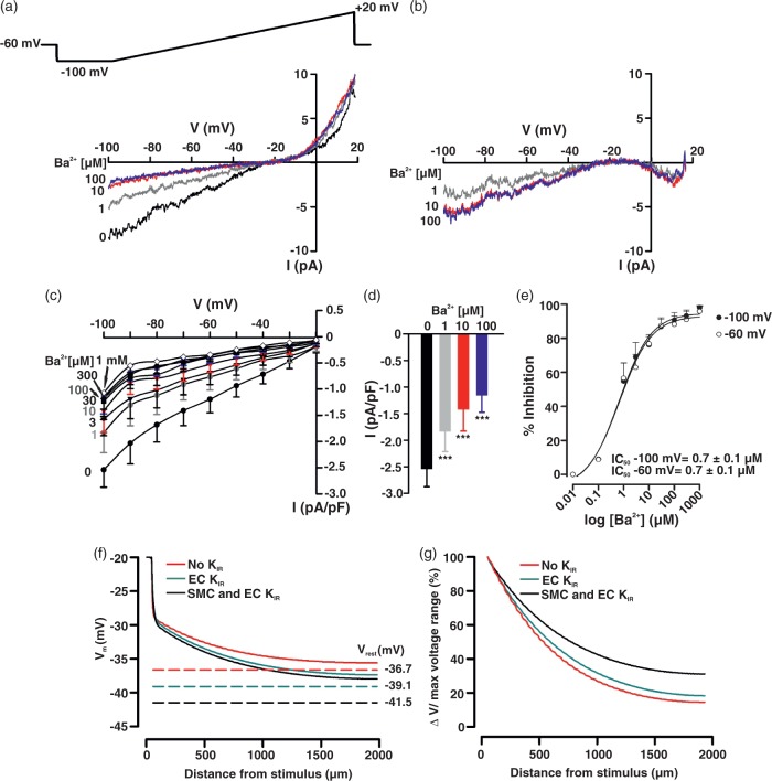
Figure 6.
KIR current in cerebral arterial endothelial cells and its virtual impact on conducted depolarization. Whole-cell K+ current was measured in isolated endothelial cells in a high K+ solution (60 mM) using a voltage ramp protocol (−100 to +20 mV) in presence and absence of BaCl2 (1 µM–1 mM). Representative whole-cell current recording (a) and Ba2+-subtracted currents (b). Summary plots (n = 7) illustrating the effect of Ba2+ (1 µM–1 mM) on whole-cell current (c) and on peak inward current at −100 mV (d). Ba2+ induces a concentration-dependent inhibition of KIR current (E, n = 7) at −100 mV (IC50 = 0.68 ± 0.02 µM) and −60 mV (IC50 = 0.67 ± 0.02 µM). Data are means ± SE. **Significant difference from control. Simulation in (f): a defined number of endothelial and smooth muscle cells in the initial 62 µm segment of the virtual vessel (see Figure 4 for details) were voltage clamped (200 ms) 20 mV positive to the resting VM (−40 mV), while voltage responses were monitored at rest (smooth muscle and endothelial KIR) and following the subtraction of KIR current in one or both cell types. Dotted lines represent resting VM (Vrest) under control conditions (black, VM: −38.7 mV), and following the subtraction of KIR conductance in the endothelium (green, VM: −39.1 mV) or both cell layers (red, VM: −36.7 mV). Conduction decay (g) along the virtual vessel under control conditions and in the absence of one or both KIR currents.

