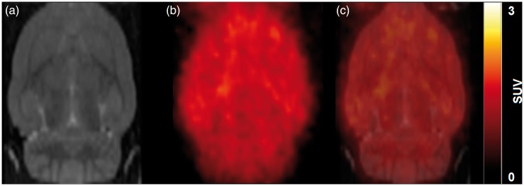
Figure 3.
[11C]Diphenhydramine brain distribution in rat. Rats are injected with [11C]diphenhydramine i.v. followed by PET acquisition for 60 min. (a) shows the rat MRI template used for delineation of the different brain regions. (b) shows a representative SUV-normalized summed [11C]diphenhydramine PET images from 0 to 60 min). Overlaid images are shown in (c).

