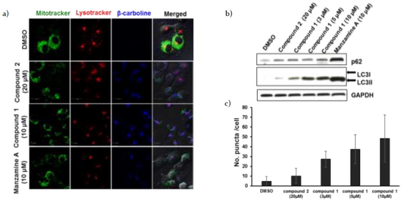
Figure 2.

a) H1299 cells were treated with compounds 1-3 for 1 hr before staining mitochondria with MitoTracker® Green FM (Invitrogen) and lysosomes with LysoTracker® Red DND-99 (Invitrogen). Confocal analysis showed localization of the β-carbolines in lysosomes. b) Western blot analysis for LC3 and p62/SQSTM1 in H1299 cells incubated with DMSO, monomer 2 (20 μM), dimer 1 (3, 5 and 10 μM) or manzamine A 3(10 μM) for 24 hrs. c) GFP-LC3 transfected H1299 cells were treated with DMSO, monomer 2 (20 μM), dimer 1 (3, 5 and 10 μM) or manzamine A 3 (10 μM) for 24 hrs. The GFP-LC3 puncta were analyzed by confocal microscope. The number of GFP-LC3 puncta/cell was scored using ImageJ software (n=30; error bars, s.d.).
