
Figure 3. Myelin repair in NG2 null mice.
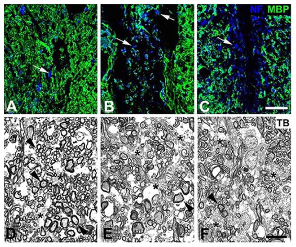
Six weeks after lysolecithin microinjection (1.5 μl of 1% lysolecithin) into the dorsal spinal cord white matter of control, OPC-NG2ko and My-NG2ko mice, animals were euthanized for determination of the extent of myelin repair. (A-C): Tissue sections were immunolabeled for myelin basic protein (MBP; green) and neurofilament protein (NF; blue), allowing visualization of myelinated axons and axons lacking association with myelin (arrows). (D-F): Toluidine blue-stained (TB) semi-thin sections enabled identification of well-myelinated axons (arrowheads) and unmyelinated axons (asterisks), along with determination of g-values for evaluating myelin thickness. Controls: A, D; OPC-NG2ko: B, E; My-NG2ko: C, F. Bar in C = 30 μm. Bar in F = 10 μm. (Reproduced with permission from Kucharova, K. and Stallcup, W. B. 2015, J. Neuroinflamm., 12, 161.)