
Figure 5. Pericyte ensheathment of endothelial cells in tumors in NG2 null mice.
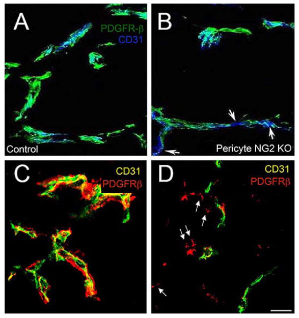
Vessels in intracranial B16F10 tumors were compared at 10 days in control (A) versus PC-NG2ko (B) mice, and in control (C) versus My-NG2ko (D) mice by immunolabeling sections for CD31 (endothelial cells) and PDGFRβ (pericytes). Pericyte (green) ensheathment of endothelial cells (blue) is incomplete in PC-NG2ko vessels (B), as judged by gaps in coverage (arrows). These gaps are not present in control vessels (A). In My-NG2ko vessels (D), many pericytes (red, arrows) fail to associate with endothelial cells (green). These detached pericytes are not seen in control vessels (C). These images also highlight the reduced diameter of vessels in My-NG2ko mice, a phenotype that is not observed in PC-NG2ko mice. Bar in D = 20 μm. (Reproduced with permission from You, W. K., Yotsumoto, F., Sakimura, K., Adams, R. H. and Stallcup, W. B. 2014, Angiogenesis, 17, 61 and from Yotsumoto, F., You, W. K., Cejudo-Martin, P., Kucharova, K., Sakimura, K. and Stallcup, W. B. 2015, Oncoimmunol., 4, e1001204.)