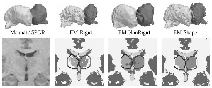
Fig. 3.
Segmentation results from different EM implementation. As clearly visible in the 2D images the shape constraint approach (EM-Shape) is closest to the expert’s segmentation indicated by the black lines. EM-Shape was also the only method who properly captured the hypothalamus (see 3D models), while EM-NonRigid is too smooth and EM-Rigid underestimated the structure.

