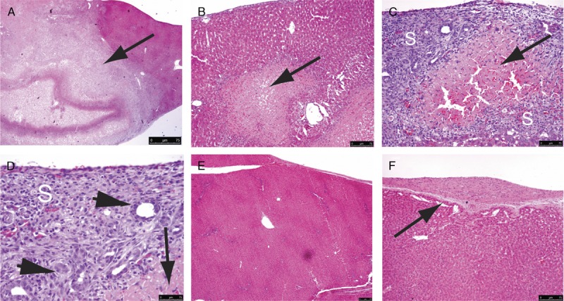
FIGURE 2.

On day 3, posttransient occlusion of the hepatic vascular inflow, geographic necrosis (A to D, arrows, magnifications ×40, ×100, ×200, and ×400, respectively) was seen in the liver parenchyma, showing IRI. The surrounding regeneration scar (S) consisted of spindle cells and ductular proliferation (C and D, arrowheads). On day 14, the liver totally regenerated with resolution of the necrosis and regeneration scars, leaving minor nodularity of architecture (E, ×100), and minor thickening of the capsule (F, arrow, ×200).
