FIGURE 5.

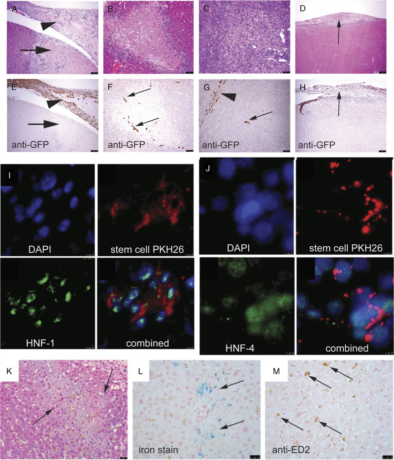
On day 3 postapplication of stem cells, most of the stem cells remained on the surface of the IR liver mixed with fibrin (A, arrowhead, ×200 and B showing immunohistochemical staining for anti-GFP). A necrotic area was seen immediately underneath the site of application (arrows). No stem cells were seen in this necrotic area. Small numbers of homed stem cells were seen at the periphery of the necrotic area (C and D, ×200). Some of the stem cells participated in the formation of the regeneration scar (E and F, ×200) At the end of day 14 buy which regeneration was completed, no GFP+ve cells were detected in the thickened fibrous tissue (G and H, arrows) or the liver parenchyma. Homed stem cells expressed HNF-1(I, ×400) and HNF-4 (J, ×400), showing differentiation toward hepatocyte lineage. When stem cells were fed with SPIO nanoparticles, these stem cells might have migrated to the periphery of necrotic area, but demised quickly, leaving the iron particles behind (K and L, arrows; H&E and iron stain, ×200, respectively). These iron particles were not bought to the necrotic area by macrophages (M, arrows, immunohistochemical staining for anti-ED2).
