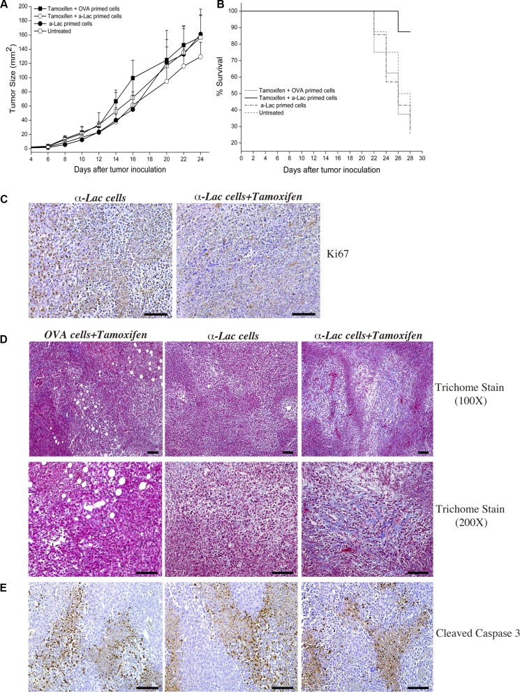
Figure 6. Amplification of target expression by ER antagonism enhances efficacy of antigen specific cell mediated immunotherapy against breast tumors.
Significant (p < 2 × 10⁻⁶) decrease in 4T1tumor progression was observed after treatment with Tamoxifen+α-Lactalbumin (α-Lac) primed lymphocytes (open circles) compared all other test groups (n = 8 each) (A). 4T1 tumors treated with α-Lactalbumin specific lymphocytes alone showed anti-tumor efficacy during initial tumor growth (closed circles), however only tumors treated with α-Lactalbumin primed lymphocytes+Tamoxifen continued to show enhanced therapeutic efficacy. Mice treated with Tamoxifen+α-Lac primed cells showed 87% survival at end of follow up at day 28 compared to 25% in controls (B; p < 0.04). Combination of α-Lac primed cell transfer with Tamoxifen diet resulted in significant reduction in cell division within 4T1 tumors as evident by Ki67 staining (C, Right Panel) compared to α-Lac specific cells alone (C, Left Panel). Tamoxifen diet+antigen specific cell transfer induced significant fibrosis (blue Trichome stain) in 4T1 tumors (D, Right Panels) compared to Tamoxifen+OVA primed cell transfer (D, Center Panels) or α-Lactalbumin primed cells alone (D, Left Panels). No significant differences in Cleaved Caspase 3 staining (brown DAB stain) were observed between treatment groups (E). Bars depict 100 μm.

