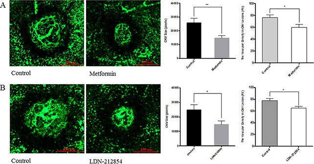
Figure 6. The effect of metformin on laser-induced choroidal neovascularization (CNV) in mice.

C57BL/6J mice were used for laser-induced CNV model. Four lesions were induced using the laser photocoagulation after anesthesia with ketamine hydrochloride. (A) Animals were equally divided into two groups (8 mice/group), treated with PBS or metformin (150 mg/kg/day) i.p. once daily from the day prior to laser irradiation (Day-1) to Day 6. For the evaluation of CNV size, at Day 7, mice were deeply anesthetized and perfused with FITC-lectin. The mice were sacrificed and the eyes were enucleated and fixed in 4% paraformaldehyde. RPE-choroid tissue was flat-mounted and observed under fluorescein microscope. Size of CNV was measured by μm2 and vascular density in CNV lesion by ImageJ program. Significance was tested by student t test (average pixels ± SEM, n=7). *p<0.05, ** p<0.01. (B) Animals were injected i.p. with PBS or LDN-212854 (6 mg/kg, twice daily) and processed as described for A. CNV size and vascular density in CNV lesions were determined. Scale bar: 200 μm.
