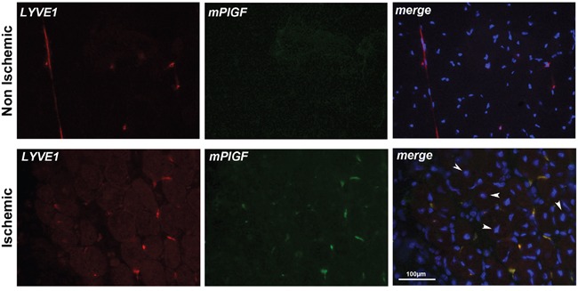
Figure 4. PlGF co-localizes with LYVE1 positive lymphatic vessels in ischemic muscle.

LYVE1 (red) identifies small inter-fiber lymphatic vessels in non-ischemic and ischemic popliteal muscles cryosections. PlGF (green) is absent in non-ischemic muscle while co-localizes with LYVE1 staining in ischemic muscle. Arrows indicate center-nucleated fibers, a characteristic of regenerating muscle that occurs in hypoxic area of muscles, that were present only in ischemic muscles. Scale bar 100μm.
